Archives for May 2011

By now you’ve likely heard something about the outbreak of food-borne illness due to E. coli that is currently causing problems in Germany. This outbreak, reportedly the world’s largest due to E. coli, has so far killed 15 people and left more than 1000 others ill.

Although the exact source of the virulent strain of this bacterium remains unknown, the pathogen has been identified on cucumbers imported from Spain. Specifically it has been identified as Shiga toxin-producing E. coli (STEC) which causes Hemolytic Uremic Syndrome (HUS).

Vegetables, however, don’t have colons for colonization with this bacterium, so how did they become “infected”?


STEC and HUS

Most research on the epidemiology of STEC in animals has involved E. coli 0157:H7, and cattle are considered the major reservoir of the STEC that result in human infection. This strain is so widely distributed among cattle that it may likely be present periodically on most farms at some stage. Although this pathogen can only survive transiently in soil when shed by affected cattle, it is, however, capable of prolonged persistence in manure and environmental waters.

Consequently, polluted groundwaters may have been the source of the bacterium in this outbreak, and the vegetables were merely vectors of disease. That is, they provided a mechanical means of transporting the STEC into people.

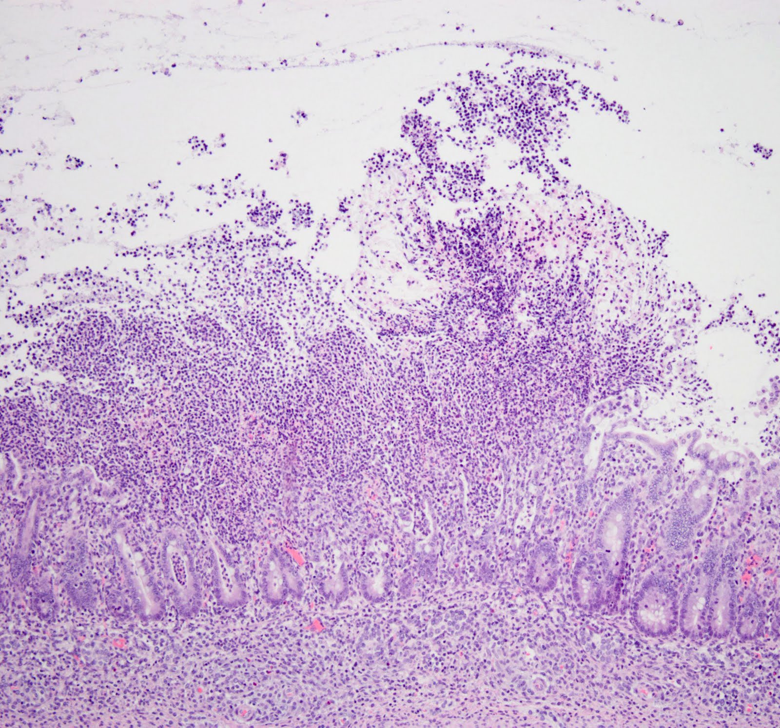
HUS typically begins with an episode of bloody diarrhea following infection of the gastrointestinal tract with E.coli 0157:H7. This pathogen produces toxins that destroy red blood cells and cause kidney damage, resulting in a combination of hemolytic anemia, thrombocytopenia and acute renal failure. HUS therefore represents a medical emergency, and although the majority of patients recover from illness, mortality rate can be up to 10%, with children and the elderley being particularly at risk.

Growing Tension

Sadly this recent STEC/HUS outbreak has additionally spread to other northern European countries, and the situation is expected to further worsen in the coming week. Subsequently, diplomatic tension between Germany and countries such as Spain, France and Russia, is rising, as some countries have banned vegetable imports from Germany and Spain.

The German public has now been warned to avoid eating raw vegetables, such as lettuce, tomatoes and cucumbers, and some products have even been removed from store shelves.

It’s been a “dog disease” kind of week on the writing front. Here’s a snapshot of it, courtesy of Wordle.


Yesterday I listened to a very interesting webinar by Richard Friedberg, MD PhD, Professor and Deputy Chairman of the Department of Pathology, Tufts University School of Medicine, Boston, MA. Dr Friedberg is also Chairman of the Pathology for Baystate Health (Springfield, MA), where he oversees a 22-pathologist academic/private practice operation responsible for 50,000 surgical specimens, 90,000 cytology specimens, 6 million laboratory tests, and a large reference laboratory.

As a pathologist myself, I had been looking forward to hearing him speak. Some of the highlights from Dr Friedberg’s webinar are outlined below:

The Evolution Of Pathology

Dr Friedberg reviewed how laboratory medicine and the practice of pathology have evolved over the decades, and how they are likely to continue to evolve in the future. Pathology has changed in numerous ways, involving a move from a formerly qualitative and later quantitative approach, to a more integrative approach currently. Additionally, our specialty has moved from “analog” to “digital”, similar to what has occurred in the field of radiology.

He talked about how, in order to thrive in highly competitive markets, clinical laboratories need to look beyond traditional models of service delivery and must now think outside the box. This may mean integrating with other diagnostic services and delivering different types of results (from laboratory to imaging) to physicians and other clients. Traditionally, “pathology” and “imaging” have been considered separate entities, but the trend is definitely moving toward a more integrated approach of diagnostic reporting that incorporates both disciplines.

Therefore, in coming years, a diagnostic report will be much more than the traditional “label” of the past. Changes including the advancement of personalized medicine, the use of electronic medical records, and the evolution of technological advancements like digital pathology, will all impact how pathologists work.  Consequently the pathologist’s report will no longer merely comprise a diagnostic name, but rather will represent an aggregate of much medical information that has been analysed using disparate tools.

The Downside Of Digitization

Some professionals have been concerned that the improvement by “moving to digital” might also have a negative effect – namely the outsourcing of pathology to overseas countries for low cost services (as has occurred in the field of radiology); however, this may not be as much of a cause for concern in pathology, for a few reasons:

Initial Image Capture: Radiological images are first obtained digitally. This is not the case with microscopic images though – the need for initial slide production will always remain. So there will always remain the need for a laboratory system to process the sample and subsequently generate the digital image from the specimen.

Image File Size: File sizes of microscopic images are much larger than radiological images, and therefore not quite as easy to forward.

Urgency: Digital forwarding of radiological images to an overseas specialist for immediate consultation has important implications in medicine. It might, for example, allow a patient in the ER to be appropriately treated or released within minutes of an imaging procedure. With surgical biopsy specimens that require routine processing, however, this urgency tends not to exist. This process is indeed still constrained by the typical turnover time of up to 24 hours or more that is required to optimally process a specimen (from formalin fixation, until a hematoxylin and eosin-stained slide is produced).   

“It’s A Mammoth”


The Tyranny Of The Tool

It is important to remember that through the centuries, pathologists have traditionally made careers out of recognizing patterns. The more recent evolution of pathology, however, means that we can no longer allow ourselves to be merely defined by our tool, the microscope. Does your job involve you working as a microscopist, or a diagnostician? Moving forward, it is most important to be the latter. We must begin to learn new ways of thinking and working in our professional environment, otherwise, allowing ourself to be defined solely by a microscope could put us out of business!

In A Nutshell

The most important thing is that we know a transition phase in pathology reporting is underway – we just don’t exactly know how long it will take. This leaves plenty of professionals currently trying to figure out where they might stand in this new, evolving healthcare approach. As pathologists, we must therefore prepare for tomorrow, and position ourselves firmly, using all the diagnostic skills we can offer in addition to microscopy.

And although the future of pathology provides a definite challenge for us all, it should, nevertheless, be a fascinating and enjoyable ride!

It’s been a really busy week on the editing and writing front and I’m looking forward to the work-free weekend. Well, at least it’ll be free of academic work!

One fun thing I discovered a couple of days ago is Wordle. You can paste in some text of your choice and watch as it generates a word cloud from them!

I thought it might be fun to try it myself and let the site generate my “week in words”! Here’s my word cloud, courtesy of some of my week’s work!

My Week In Words!

So there you have it! Why don’t you head over there yourself and see what kind of word cloud your week generates!


For the past four years I have been a PhD thesis committee member for a graduate student. Last week she gave her final thesis defense seminar, describing her research into the pathogenesis of Clostridium difficile infection (CDI). I’m pleased and proud to say that she performed wonderfully, presenting a beautiful synopsis of her research, and additionally dealt extremely well with our questions from the committee afterwards. (Congratulations Jenny!)

Interestingly, I overheard a conversation at the checkout in my local pharmacy just an hour later. One gentleman was telling the cashier how his wife had recently been taking antibiotics in hospital, and had subsequently developed a terrible diarrhea-related illness.

Needless to say, it turned out that she had CDI. Quite bizarre that I had just spent a whole afternoon discussing this very disease! So it seems like a fitting opportunity to briefly mention this important condition.

Clostridium difficile

Clostridium difficile is a gram positive, spore-forming, anaerobic, toxin-producing bacterial organism. Two of its toxins in particular, TcdA and TcdB, are responsible for most of the important, pathologic changes associated with its infection.

Clostridium difficile Infection (CDI)

This agent causes a spectrum of intestinal pathology in people that ranges from diarrhea to life-threatening necrotizing colitis.  The incidence of CDI has certainly risen dramatically over the past 30 years or so, specifically in association with increasing use of antibiotics in healthcare. Interestingly, CDI is the most common infection acquired by hospitalized patients, and its  incidence seems to have surpassed that of Methicillin-resistant Staphylococcus aureus (MRSA) infections in hospitals. Although elderly patients are at greatest risk of infection, the incidence of disease in younger patients is actually increasing.

Pathophysiology

The inciting event for Clostridium difficile-associated disease is disruption of the normal microflora of the bowel – typically due to broad spectrum antibiotic therapy. Clostridium difficile spores can persist in the environment, and are ingested by patients. Typically they cause no problem, but following disruption of the bowel’s microflora in these antibiotic-treated patients, their ingestion subsequently allows Clostridium difficile to colonize the colon. Host-associated factors (such as age, immune status, concurrent disease status) now determine what happens next.

Either a patient will become an asymptomatic carrier, or can develop disease. This disease can range from mild diarrhea to potentially fatal pseudomembranous colitis. This colitis arises as a result of the action of toxins TcdA and TcdB.

TcdA especially seems to have enterotoxic activity. It activates inflammatory cells, stimulating them to produce inflammatory mediators that lead to fluid secretion and increased intestinal mucosal permeability. As inflammation progresses, the colonic mucosa becomes ulcerated, with accumulation of inflammatory cells and necrotic cell debris on the colonic surface. This produces the classic pseudomembrane overlying the mucosal surface. Both TcdA and TcdB additionally have cytotoxic activity that contributes to the colitis of CDI.

Most cases of CDI are known to occur between 4-9 days of antibiotic therapy, and a patient’s risk of developing diarrhea due to CDI doubles with more than 3 days of antibiotic therapy. This organism is responsible for approximately 25% of cases of antibiotic-associated diarrhea, and up to 75% of cases of antibiotic-associated colitis in people. Current estimates suggest that there are approximately 500,000 cases of CDI in the US annually, and up to 20,000 of these cases result in death.

Severe inflammation and ulceration of the colon
due to Clostridium difficile infection


So clearly CDI is an important problem amongst hospitalized patients. Its increasing incidence, combined with the associated financial implications to the healthcare system, certainly contribute to the fact that this condition is being widely researched in the scientific community.

A heart attack in the morning may be more serious than one at any other time of day.

This finding was reported in a research article published online ahead of print, in the journal Heart.     


Background:

It is known that the 24 hour body clock influences cardiovascular physiological processes. It also influences the incidence of cardiac arrests, which tend to occur more frequently when a person is waking up from sleep. What is not fully known, however, is how the time of occurrence of the heart attack influences the extent of damage to the heart.

A group of Spanish researchers evaluated data from 811 patients with a STEMI cardiac arrest who were admitted to a coronary care unit in Madrid between 2003 and 2009. A “STEMI” heart attack is an “ST segment elevation myocardial infarction” – a type of heart attack that results from blockage of blood supply to the heart over a prolonged period of time. The group aimed to assess whether time of day of a heart attack affected the size of infarct (dead cardiac tissue), and they determined infarct size by evaluating enzyme release in patients. Patients were grouped according to STEMI onset time, into one of four 6-hour time periods (in phase with 24-hour body clock rhythms.)


Results

Patients who had a heart attack between 6am and noon (in the “dark to light” transition period) had the largest infarct sizes, as well as a 21% higher level of enzymes during this period (indicative of a larger infarct size) than patients who experienced a heart attack between 6pm and midnight.

The majority of patients (269) had their heart attack between 6am and noon. Of the remaining patients, 240 had their attack between noon and 6pm, 161 had theirs between 6pm and midnight, and 141 had theirs between midnight and 6am. Patients with a STEMI in the anterior wall of the heart also developed a larger infarct than patients who had a heart attack in other cardiac locations.


Conclusion:

Heart attacks occurring between 6am and noon are likely to produce a 20% larger infarct (which is inevitably more serious for the patient) than at any other time of the day.

The authors subsequently concluded: “If confirmed, these results may have a significant impact on the interpretation of clinical trials of cardioprotective strategies in STEMI.”


Circadian Variations Of Infarct Size In Acute Myocardial Infarction. (2011). Heart, Apr 27 (Epub ahead of print). Suarez-Barrientos et al.

Academia can be a “publish or perish” environment at the best of times – so after working long, hard hours to collect all your scientific data, you deserve a final article that is free of errors and most accurately represents your efforts. You may have extremely impressive scientific data, but if your manuscript lacks readability and clarity, it will typically be rejected by the peer-review process when submitted for publication.


Language and Publication in “Cardiovascular Research” Articles [Cardiovasc Res (2002) Feb 1:53(2):279-285] by Coates et al, surveyed 120 articles that were submitted to the journal “Cardiovascular Research”. They reported: “There is a clear indication that badly written articles correlated with a high rejection rate.”  Additionally they stated: “On equal scientific merit, a badly written article will have less chance of being accepted … even if the editor does not identify language as a motive for rejection.”   

So the way in which your data is communicated is almost as crucial as the data itself. Writing quality clearly influences whether a paper is accepted for publication, regardless of the research quality. As with all kinds of writing, it is therefore extremely valuable to allow a fresh pair of eyes to review your finished paper – a skilled academic editor will not only proofread your work, but will greatly enhance its scientific clarity. This will therefore not only improve its chances of being published, but also speed up the publication time.

Writing therefore plays a pivotal role in the communication of your scientific data. Therefore, although the process of editing and revising your manuscript can seem overwhelming (especially if English is not your native language), it is a necessary step if you hope to maximize the impact of your work within the scientific community.