Archives for June 2011

A recent study indicates that being married might actually improve the likelihood of survival for patients with colon cancer.

Researchers studied the medical records of 127, 753 patients and determined that married people with colon cancer were 14% less likely to die than unmarried patients with the disease. Interestingly, the benefit of marriage was almost identical in both men and women. The study also found that married patients were typically diagnosed at earlier stages of colon cancer, and opted for more aggressive treatment – similar to findings from studies of other types of cancer.

Although the reason for these findings isn’t totally clear, the researchers suggest that the support and caregiving from spouses may result in improved cancer management, and better disease outcomes as a result.

Colon cancer is the 3rd most common type of cancer in the USA, and represents a leading cause of cancer-related deaths. The death rate from this type of cancer, however, has been decreasing over the past 20 years. One reason for this is likely the increased use of screening techniques to diagnose colon cancer – this in turn allows polyps to be detected and removed earlier before they can become cancerous. It also allows colon cancers to be detected at earlier stages when the disease is actually easier to treat and potentially cure.

Consequently there are currently over one million survivors of colorectal cancer in the USA.

Li Wang et al. Marital status and colon cancer outcomes in US Surveillance, Epidemiology and End Results registries: Does marriage affect cancer survival by gender and stage? Cancer Epidemiology, 2011 DOI: http://bit.ly/ig0pCz

Welcome to my new pad! I’m in the process of switching over here to WordPress, so please bear with me. I’m having some issues with the format of imported posts, so previous posts may look a little odd, but hopefully the change will be a good one overall.

I’ve just finished editing a huge hematology review article on erythropoietin, so it seemed fitting to create this week’s Wordle cloud from the work I did on that. 

Exposure To Pets In The First Year Of Life May Reduce Risk Of Allergies

The relationship between exposure to pets and allergic disease risk has long been unresolved. Various studies in recent years have examined the effect of early exposure to cats and dogs on allergies in children. The results, however, have been conflicting, with some showing a benefit from having a pet, while others indicated that it may actually make children more prone to developing allergies.

The focus of the study was to evaluate the association between lifetime dog and cat exposure and allergic sensitization to the specific animal at 18 years of age. Researchers collected information from 566 children and their parents about the childrens’ exposure to indoor pets, and their history of allergies.

A study published online ahead of print in the journal “Clinical & Experimental Allergy”, however, suggests that having a pet in the house during the first year of a child’s life could halve the risk of them becoming allergic to animals [“Lifetime Dog and Cat Exposure and Dog- and Cat-Specific Sensitization at Age 18 Years.” Wegienka et al, 2011: 41(7)979-986.]

They followed the children from birth to 18 years old and found that exposure to a cat in the first year of life was associated with a halved risk of having an immune system that was sensitized to cat allergens. The findings for dogs were more complicated, however, and a link between exposure and reduced risk of sensitization later on was found in boys only.

The results of this study now suggest that researchers should further study the first year of life, since this may be the critical window during childhood when indoor exposure to pets may influence sensitization to them.

“This research provides further evidence that experiences in the first year of life are associated with health status later in life, and that early life pet exposure does not put most children at risk of being sensitized to these animals later in life,” said first author Ganesa Wegienka.

Yesterday I went to the New England Chapter meeting of the American Medical Writers Association. It was a lot of fun, highly informative & I met some great new people. If you’re a science writer but are not yet a member of the AMWA, I’d highly recommend it – a great networking opportunity.


Since I’m a veterinary pathologist by training, I thought you might like me to share some aspects of this side of my work with you. 

Some of my daily routine involves microscopic evaluation of sections of tissue from animals, often for diagnostic purposes. So I’ll try and share some interesting cases with you from time to time.

I’ll start off today by sharing information about a case of herpesvirus infection in a cat.


History: A two week old kitten died following four days of respiratory distress, with pronounced sneezing and coughing.

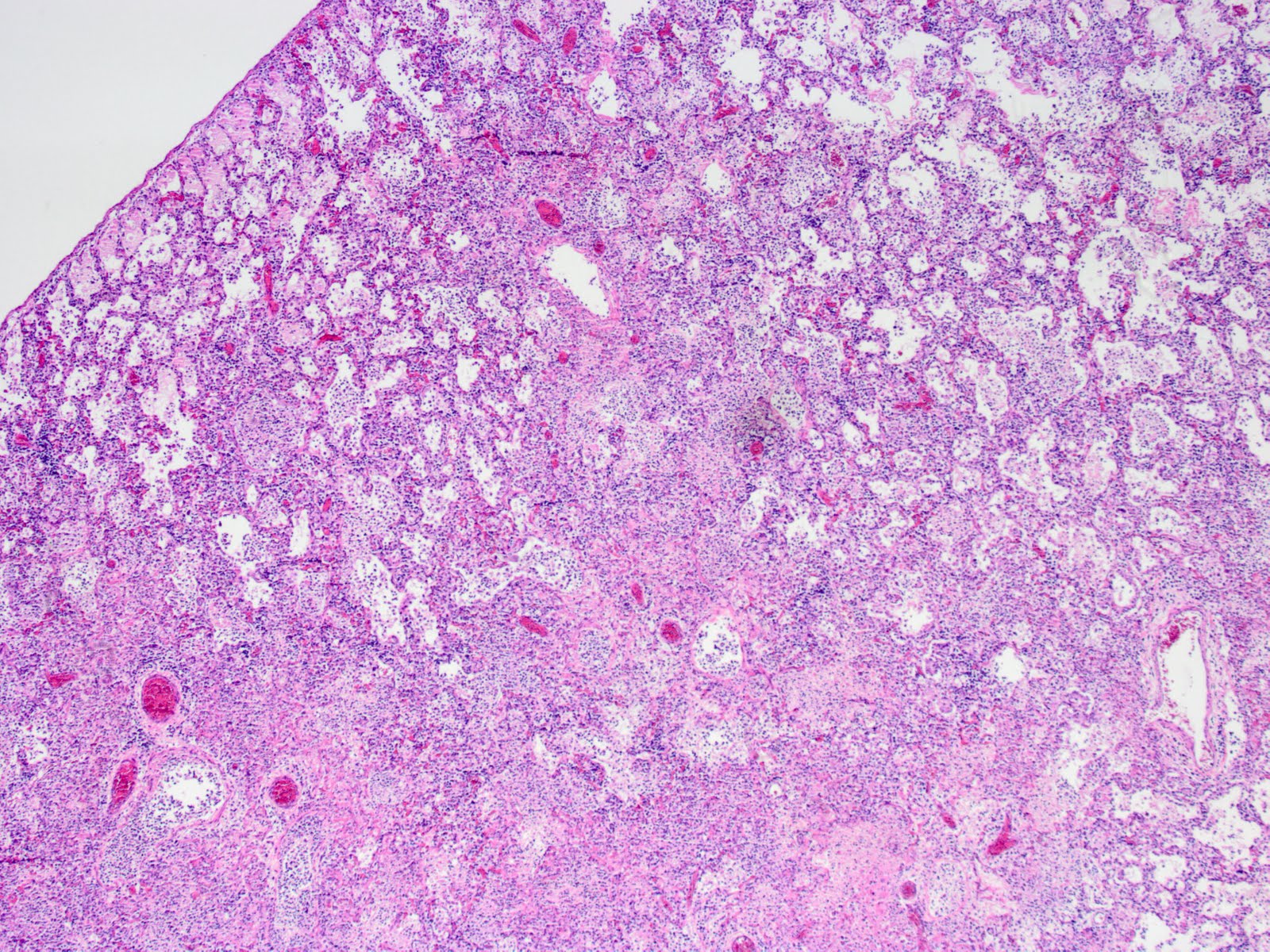
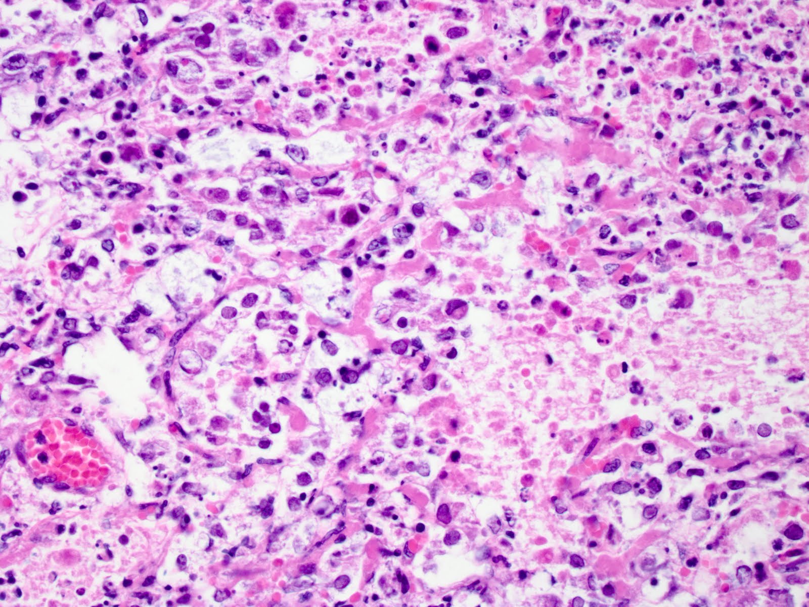
Diagnosis: A diagnosis of pneumonia due to FHV-1 (feline herpesvirus-1) infection was made after microscopic evaluation of tissues taken at postmortem examination. 

FHV-1 IN CATS: This is a common feline virus that is transmitted between cats by direct contact and exchange of nasal and ocular fluids. “Feline rhinotracheitis” is the classic condition associated with this virus, and typically manifests as upper respiratory disease and conjunctivitis in kittens, similar to this case. Systemic illness with interstitial pneumonia has also been reported in kittens, and such cases of systemic disease can arise as a result of immunosuppression. Additionally, the virus can cause conjunctivitis as well as facial and nasal dermatitis or stomatitis in adult cats.

The usually clear air spaces in the lung are
filled with inflammatory cells & debris
Higher magnification view showing intense inflammation 
with necrotic tissue and debris, along with the characteristic 
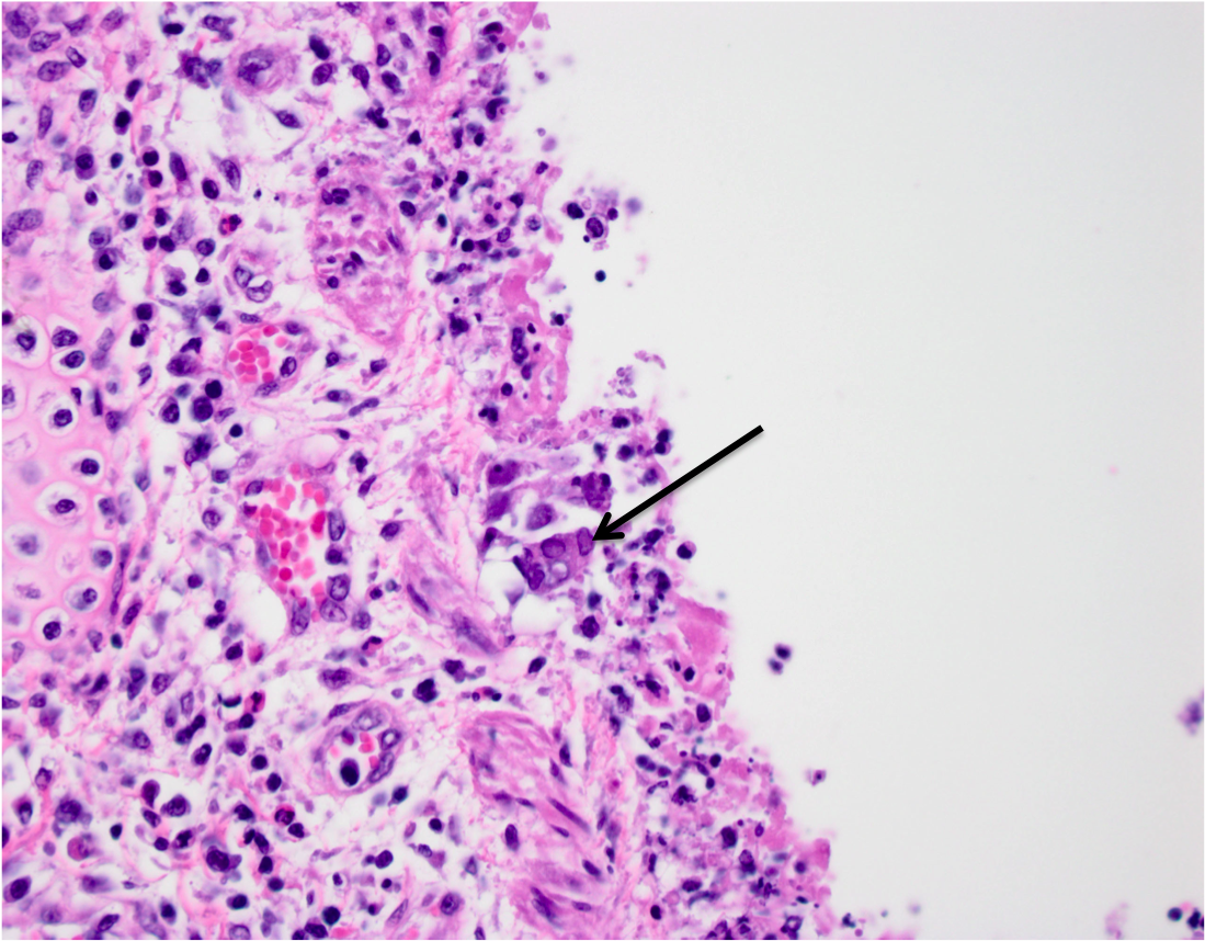
diagnostic finding of viral inclusion bodies in cells. 
The arrow points to a syncitial cell containing multiple nuclei.
These nuclei contain characteristic viral inclusion bodies


Although this is a devastating disease, the good news is that cats can be vaccinated against the causative virus, thus reducing the incidence or severity of disease.

Cats can be carriers of FHV-1 though – in such cases, if a female cat carrying the virus is infected before she is vaccinated, the vaccination will not be effective in eliminating the latent infection. Subsequently, any stress, such as that associated with pregnancy, will result in viral recrudescence and shedding. Following birth, the mother cat will transmit the virus to her kittens in her milk, or through grooming, direct contact or nasal secretions.

So if your cat or kitten has any signs of respiratory disease – be sure to have him or her examined by your veterinarian so as to ensure the correct diagnosis can be made, and appropriate treatment can follow.

This week I devoted some time to updating my CV, so I thought I’d see how that turned out on Wordle.

On this day in 1944, Allied Forces stormed the beaches of Normandy, France marking what would become forever remembered as the D-Day invasion of World War II. 


Exact statistics vary across different resources, but some 300,000+ American service members were killed during WWII, and an additional 671,000+ were wounded in action. Since September 11th, 2001, 5000+ service members have been killed, and more than 36,000 have been injured.

In the decades that have spanned the end of WWII and present times, significant advancements have not only been military, but also medical. In tribute to all who have served their countries in wars throughout the world, the American Academy of Orthopaedic Surgeons has organized “Wounded in Action: An Art Exhibition of Orthopaedic Advancements“.

Currently on display at the Chicago Cultural Center, this exhibition honors the sacrifices made by injured military service members, and explores the emotional aspects of battle injury. Paintings, prints, photographs and sculptures are amongst the 107 pieces of work on display from 77 contributors. Contributing artists include military service members, family members and military surgeons who have cared for the wounded.

If you are fortunate enough to be able to visit this exhibition you’ll see work from Sergeant Pete Damon. Sergeant Damon is an Iraq War veteran who lost parts of both arms in 2003. His artwork is absolutely fantastic and I thoroughly recommend you to at least check out his work on his blog. I’ve seen quite a bit of it, he’s from here in Massachusetts where I live. His story and his talent are extremely inspirational……

…….To put it mildly.

This week has involved our group finalizing numerous research abstracts that need to be submitted ready for this year’s AALAS meeting. So it’s been a very intestinal-oriented week. With somewhat of a helicobacter bias!